
Capturing adhesion molecules in action through imaging
ApplyProject Description
Cell adhesion occurs through spatio-temporally regulated interactions that are mediated by multiple intra- and inter-cellular components. Physiologically, shear forces on flowing cells orchestrate these interactions. The conventional assay used to study the effect of shear flow on cell adhesion is the parallel plate flow chamber (PPFC) assay, which records videos of cells rolling in flow and adhering to adhesion molecules on cells (eg. endothelial) or immobilized to a surface. However, due to the limited spatial resolution and sensitivity of these assays, nanoscopic molecular level mechanisms of selectin-selectin ligand interactions and their role in leukocyte (neutrophils, HSCs, T-cells) migration can’t be assessed.
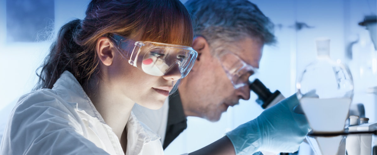
Recent developments in super resolution and single-molecule fluorescence imaging techniques allow for the visualization of individual molecules with nanometer spatial resolution and millisecond temporal resolution. Furthermore, advanced super-resolution microscopy provides unique opportunities to obtain information about nanometer-scale conformational dynamics of protein complexes as well as nanoscale architectures of biological samples. In collaboration with S. Habuchi (BESE, KAUST), whose research focuses on the development of tools and materials for fluorescence molecular imaging, we optimized the PPFC assay to image the cell using super resolution fluorescence microscopy.
This allows us to image single molecular ligand architecture on the cell surface and determine how the ligand distribution was influenced as a result of rolling on selectin (E-/P-selectin) surfaces, both to each other (clustering) and to other selectin ligands. To characterize the dynamics of ligand distribution, we also developed real-time live cell imaging of these ligands under shear flow and are able to beautifully observe long, thin, flexible structures protruding out from the rear (tethers) and the front (slings) sides of the cell as it rolls over selectin expressing surfaces.
For this project, these novel-imaging tools, combined with molecular, cellular and proteomic technologies will be used to further understand the cellular landscape that results before, during and following the migration of model cells such as hematopoietic stem cells.
 Division -
Biological and Environmental Sciences and Engineering
Division -
Biological and Environmental Sciences and Engineering
About the
Researcher
Jasmeen Merzaban

Desired Project Deliverables
- overall goal is to develop a platform for imaging cells in flow over tissue samples expressing selectins
- growth and maintenance of cell lines and primary cells
- prepare and stain tissue sections for selectins (E-/P-/L-selectin)
- prepare and stain cells lines for selectin ligands
- run flow assays and use super resolution imaging under the supervision of an experienced PhD student